The Stanford Digital Brain Project
How AI could help predict human brain activity
Page 1
BUNCOSMOS RESEARCH LAB RESEARCH DEEP DIVE / 04 NOV 2026
The Stanford Digital Brain Project
How AI could help predict human brain activity
Conceptual illustration - not a brain scan
The experiments, the imaging, the scientific precedents
and the evidence still needed
buncosmos.ai Public evidence reviewed through 4 November 2026
Page 2
BUNCOSMOS RESEARCH LAB AI AND NEUROSCIENCE / 04 NOV 2026
RESEARCH BRIEF
The project at a glance
Stanford has launched a five-year effort to learn predictive models of human brain
activity from carefully designed experiments. The immediate deliverable is a
scientific resource: measurements, reusable models and tests of their predictions.
The difficult question is whether those predictions will also reveal how the brain
works.
What is established
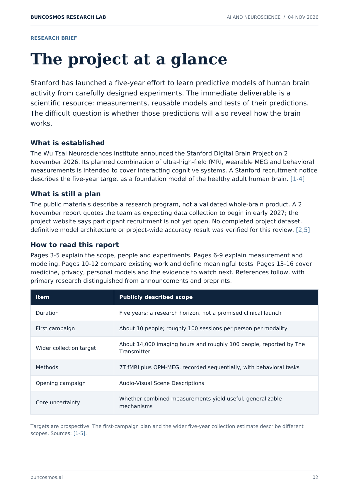
The Wu Tsai Neurosciences Institute announced the Stanford Digital Brain Project on 2 November 2026. Its planned combination of ultra-high-field fMRI, wearable MEG and behavioral measurements is intended to cover interacting cognitive systems. A Stanford recruitment notice describes the five-year target as a foundation model of the healthy adult human brain. [1-4]
What is still a plan
The public materials describe a research program, not a validated whole-brain product. A 2 November report quotes the team as expecting data collection to begin in early 2027; the project website says participant recruitment is not yet open. No completed project dataset, definitive model architecture or project-wide accuracy result was verified for this review. [2,5]
How to read this report
Pages 3-5 explain the scope, people and experiments. Pages 6-9 explain measurement and modeling. Pages 10-12 compare existing work and define meaningful tests. Pages 13-16 cover medicine, privacy, personal models and the evidence to watch next. References follow, with primary research distinguished from announcements and preprints.
Item Publicly described scope
Duration Five years; a research horizon, not a promised clinical launch
First campaign About 10 people; roughly 100 sessions per person per modality
Wider collection target About 14,000 imaging hours and roughly 100 people, reported by The Transmitter
Methods 7T fMRI plus OPM-MEG, recorded sequentially, with behavioral tasks
Opening campaign Audio-Visual Scene Descriptions
Core uncertainty Whether combined measurements yield useful, generalizable mechanisms
Targets are prospective. The first-campaign plan and the wider five-year collection estimate describe different scopes. Sources: [1-5].
buncosmos.ai 02
Page 3
BUNCOSMOS RESEARCH LAB AI AND NEUROSCIENCE / 04 NOV 2026
01 / SCIENTIFIC SCOPE
What a large brain model would predict
A model of responses and dynamics
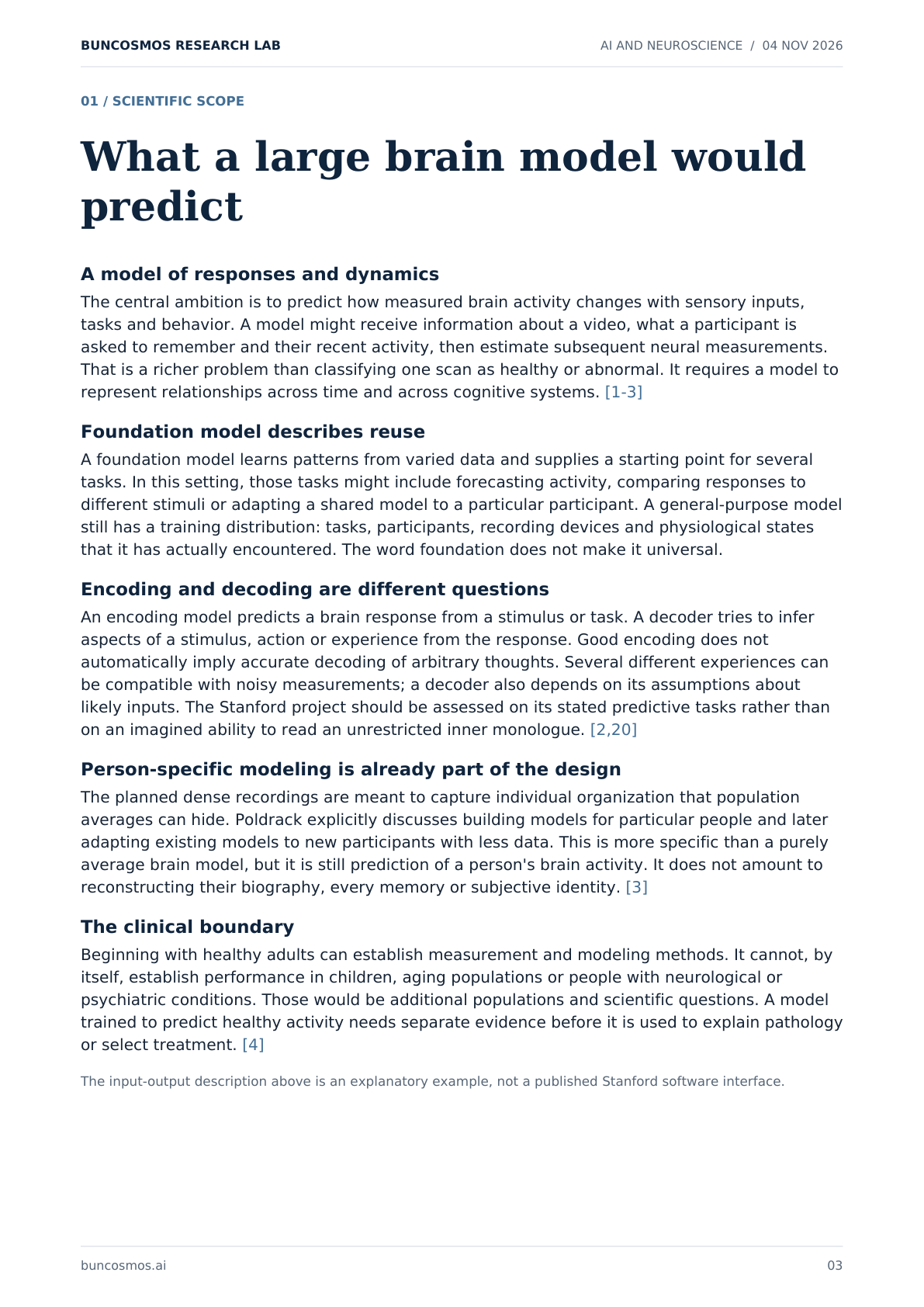
The central ambition is to predict how measured brain activity changes with sensory inputs, tasks and behavior. A model might receive information about a video, what a participant is asked to remember and their recent activity, then estimate subsequent neural measurements. That is a richer problem than classifying one scan as healthy or abnormal. It requires a model to represent relationships across time and across cognitive systems. [1-3]
Foundation model describes reuse
A foundation model learns patterns from varied data and supplies a starting point for several tasks. In this setting, those tasks might include forecasting activity, comparing responses to different stimuli or adapting a shared model to a particular participant. A general-purpose model still has a training distribution: tasks, participants, recording devices and physiological states that it has actually encountered. The word foundation does not make it universal.
Encoding and decoding are different questions
An encoding model predicts a brain response from a stimulus or task. A decoder tries to infer aspects of a stimulus, action or experience from the response. Good encoding does not automatically imply accurate decoding of arbitrary thoughts. Several different experiences can be compatible with noisy measurements; a decoder also depends on its assumptions about likely inputs. The Stanford project should be assessed on its stated predictive tasks rather than on an imagined ability to read an unrestricted inner monologue. [2,20]
Person-specific modeling is already part of the design
The planned dense recordings are meant to capture individual organization that population averages can hide. Poldrack explicitly discusses building models for particular people and later adapting existing models to new participants with less data. This is more specific than a purely average brain model, but it is still prediction of a person's brain activity. It does not amount to reconstructing their biography, every memory or subjective identity. [3]
The clinical boundary
Beginning with healthy adults can establish measurement and modeling methods. It cannot, by itself, establish performance in children, aging populations or people with neurological or psychiatric conditions. Those would be additional populations and scientific questions. A model trained to predict healthy activity needs separate evidence before it is used to explain pathology or select treatment. [4]
The input-output description above is an explanatory example, not a published Stanford software interface.
buncosmos.ai 03
Page 4
BUNCOSMOS RESEARCH LAB AI AND NEUROSCIENCE / 04 NOV 2026
02 / PEOPLE AND SUPPORT
A consortium with two kinds of expertise
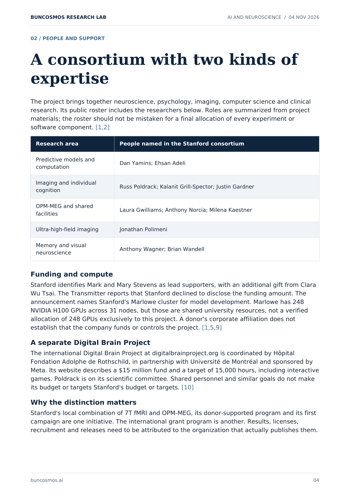
The project brings together neuroscience, psychology, imaging, computer science and clinical research. Its public roster includes the researchers below. Roles are summarized from project materials; the roster should not be mistaken for a final allocation of every experiment or software component. [1,2]
Research area People named in the Stanford consortium
Predictive models and computation Dan Yamins; Ehsan Adeli
Imaging and individual cognition Russ Poldrack; Kalanit Grill-Spector; Justin Gardner
OPM-MEG and shared facilities Laura Gwilliams; Anthony Norcia; Milena Kaestner
Ultra-high-field imaging Jonathan Polimeni
Memory and visual neuroscience Anthony Wagner; Brian Wandell
Funding and compute
Stanford identifies Mark and Mary Stevens as lead supporters, with an additional gift from Clara Wu Tsai. The Transmitter reports that Stanford declined to disclose the funding amount. The announcement names Stanford's Marlowe cluster for model development. Marlowe has 248 NVIDIA H100 GPUs across 31 nodes, but those are shared university resources, not a verified allocation of 248 GPUs exclusively to this project. A donor's corporate affiliation does not establish that the company funds or controls the project. [1,5,9]
A separate Digital Brain Project
The international Digital Brain Project at digitalbrainproject.org is coordinated by Hôpital Fondation Adolphe de Rothschild, in partnership with Université de Montréal and sponsored by Meta. Its website describes a $15 million fund and a target of 15,000 hours, including interactive games. Poldrack is on its scientific committee. Shared personnel and similar goals do not make its budget or targets Stanford's budget or targets. [10]
Why the distinction matters
Stanford's local combination of 7T fMRI and OPM-MEG, its donor-supported program and its first campaign are one initiative. The international grant program is another. Results, licenses, recruitment and releases need to be attributed to the organization that actually publishes them.
buncosmos.ai 04
Page 5
BUNCOSMOS RESEARCH LAB AI AND NEUROSCIENCE / 04 NOV 2026
03 / THE FIRST CAMPAIGN
Recording the same people in depth
ONE SCENE
ATTEND
REMEMBER
DESCRIBE
Focus on a feature
Recognize an event
Put it into words
Video + audio
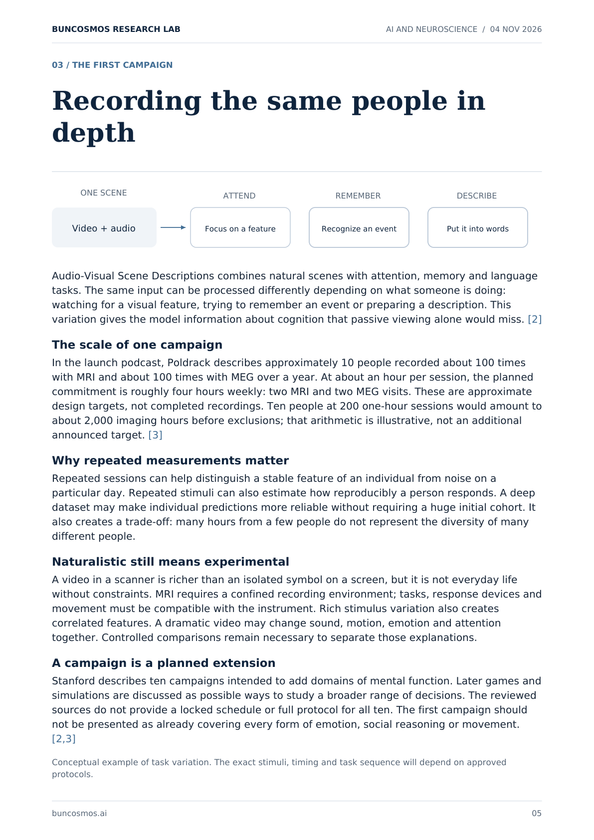
Audio-Visual Scene Descriptions combines natural scenes with attention, memory and language tasks. The same input can be processed differently depending on what someone is doing: watching for a visual feature, trying to remember an event or preparing a description. This variation gives the model information about cognition that passive viewing alone would miss. [2]
The scale of one campaign
In the launch podcast, Poldrack describes approximately 10 people recorded about 100 times with MRI and about 100 times with MEG over a year. At about an hour per session, the planned commitment is roughly four hours weekly: two MRI and two MEG visits. These are approximate design targets, not completed recordings. Ten people at 200 one-hour sessions would amount to about 2,000 imaging hours before exclusions; that arithmetic is illustrative, not an additional announced target. [3]
Why repeated measurements matter
Repeated sessions can help distinguish a stable feature of an individual from noise on a particular day. Repeated stimuli can also estimate how reproducibly a person responds. A deep dataset may make individual predictions more reliable without requiring a huge initial cohort. It also creates a trade-off: many hours from a few people do not represent the diversity of many different people.
Naturalistic still means experimental
A video in a scanner is richer than an isolated symbol on a screen, but it is not everyday life without constraints. MRI requires a confined recording environment; tasks, response devices and movement must be compatible with the instrument. Rich stimulus variation also creates correlated features. A dramatic video may change sound, motion, emotion and attention together. Controlled comparisons remain necessary to separate those explanations.
A campaign is a planned extension
Stanford describes ten campaigns intended to add domains of mental function. Later games and simulations are discussed as possible ways to study a broader range of decisions. The reviewed sources do not provide a locked schedule or full protocol for all ten. The first campaign should not be presented as already covering every form of emotion, social reasoning or movement. [2,3]
Conceptual example of task variation. The exact stimuli, timing and task sequence will depend on approved protocols.
buncosmos.ai 05
Page 6
BUNCOSMOS RESEARCH LAB AI AND NEUROSCIENCE / 04 NOV 2026
04 / THE SPATIAL MEASUREMENT
What 7T fMRI can actually see
Neural processing
BOLD response
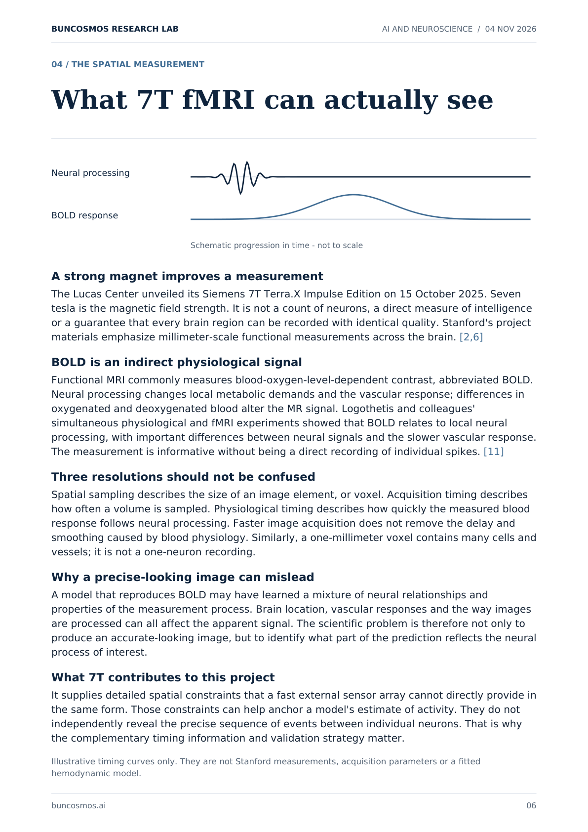
Schematic progression in time - not to scale
A strong magnet improves a measurement
The Lucas Center unveiled its Siemens 7T Terra.X Impulse Edition on 15 October 2025. Seven tesla is the magnetic field strength. It is not a count of neurons, a direct measure of intelligence or a guarantee that every brain region can be recorded with identical quality. Stanford's project materials emphasize millimeter-scale functional measurements across the brain. [2,6]
BOLD is an indirect physiological signal
Functional MRI commonly measures blood-oxygen-level-dependent contrast, abbreviated BOLD. Neural processing changes local metabolic demands and the vascular response; differences in oxygenated and deoxygenated blood alter the MR signal. Logothetis and colleagues' simultaneous physiological and fMRI experiments showed that BOLD relates to local neural processing, with important differences between neural signals and the slower vascular response. The measurement is informative without being a direct recording of individual spikes. [11]
Three resolutions should not be confused
Spatial sampling describes the size of an image element, or voxel. Acquisition timing describes how often a volume is sampled. Physiological timing describes how quickly the measured blood response follows neural processing. Faster image acquisition does not remove the delay and smoothing caused by blood physiology. Similarly, a one-millimeter voxel contains many cells and vessels; it is not a one-neuron recording.
Why a precise-looking image can mislead
A model that reproduces BOLD may have learned a mixture of neural relationships and properties of the measurement process. Brain location, vascular responses and the way images are processed can all affect the apparent signal. The scientific problem is therefore not only to produce an accurate-looking image, but to identify what part of the prediction reflects the neural process of interest.
What 7T contributes to this project
It supplies detailed spatial constraints that a fast external sensor array cannot directly provide in the same form. Those constraints can help anchor a model's estimate of activity. They do not independently reveal the precise sequence of events between individual neurons. That is why the complementary timing information and validation strategy matter.
Illustrative timing curves only. They are not Stanford measurements, acquisition parameters or a fitted hemodynamic model.
buncosmos.ai 06
Page 7
BUNCOSMOS RESEARCH LAB AI AND NEUROSCIENCE / 04 NOV 2026
05 / THE TEMPORAL MEASUREMENT
What wearable MEG adds
Magnetic fields from coordinated neural currents
MEG detects magnetic fields associated with brain activity. Optically pumped magnetometers use atomic properties of alkali metals to sense extremely weak fields. A 2018 Nature study demonstrated a wearable OPM system during movement, establishing an important predecessor to today's systems. This is quantum sensing, not a quantum computer operating inside the participant's head. [7,12]
Stanford's installed system
Stanford's September 2026 description identifies 144 sensors at the Koret Human Neurosciences Community Laboratory. Sensors close to the scalp and adaptable positioning support more natural movement than a conventional fixed cryogenic MEG helmet. A magnetically shielded room and carefully designed stimulus equipment are necessary because environmental magnetic fields can overwhelm the brain signals. Wearable does not mean the current experiment can take place anywhere without shielding. [7]
Fast signals still require spatial inference
The sensor array measures a magnetic field outside the head. Researchers must infer the internal sources that could have generated it. Different source configurations may explain similar external fields, making anatomy, geometry and modeling assumptions important. Millisecond timing is a strength; it does not mean that each sensor has uniquely identified a particular neuron.
Where and when is a useful shorthand
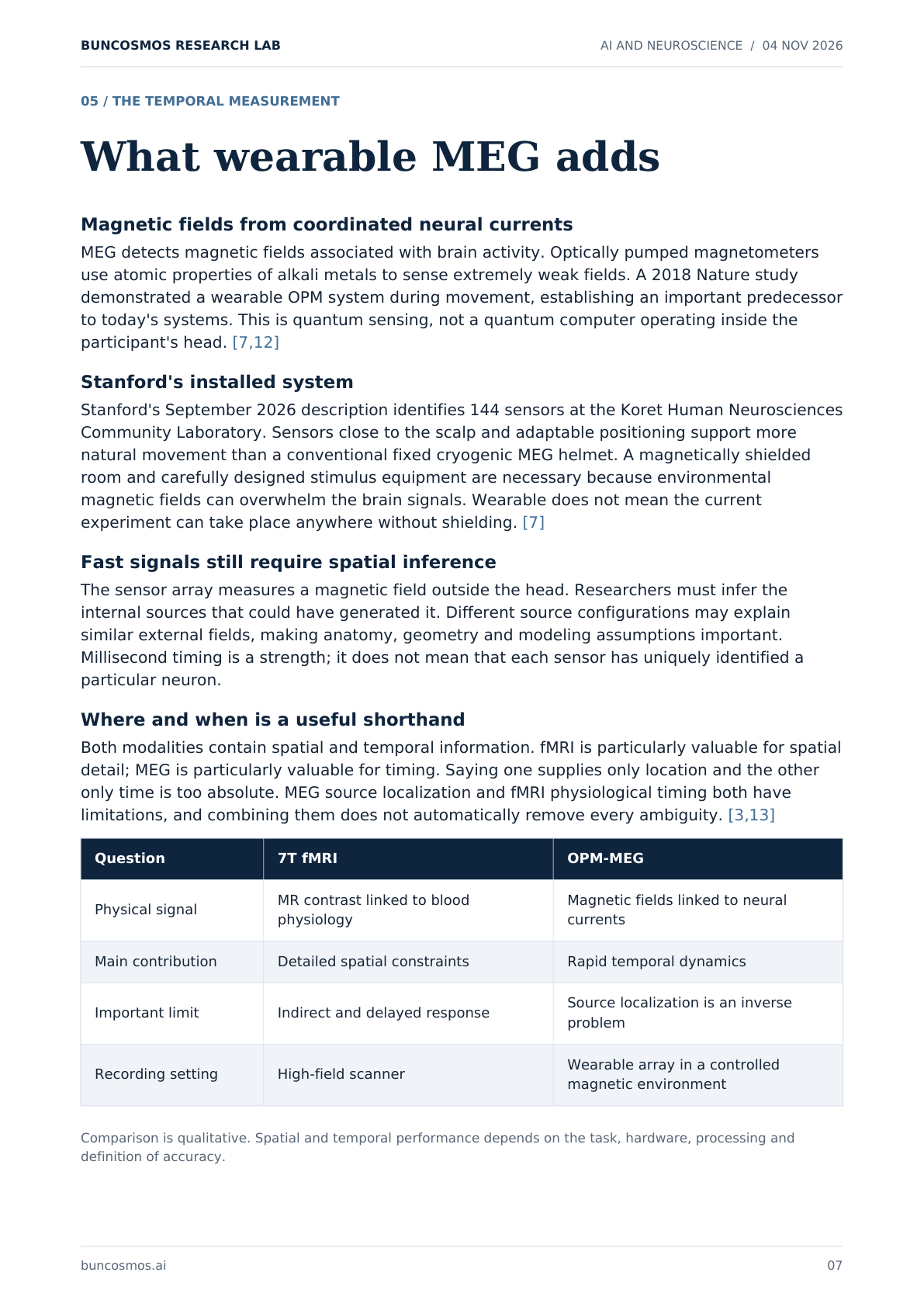
Both modalities contain spatial and temporal information. fMRI is particularly valuable for spatial detail; MEG is particularly valuable for timing. Saying one supplies only location and the other only time is too absolute. MEG source localization and fMRI physiological timing both have limitations, and combining them does not automatically remove every ambiguity. [3,13]
Question 7T fMRI OPM-MEG
Physical signal MR contrast linked to blood physiology
Magnetic fields linked to neural currents
Main contribution Detailed spatial constraints Rapid temporal dynamics
Important limit Indirect and delayed response Source localization is an inverse problem
Recording setting High-field scanner Wearable array in a controlled magnetic environment
Comparison is qualitative. Spatial and temporal performance depends on the task, hardware, processing and definition of accuracy.
buncosmos.ai 07
Page 8
BUNCOSMOS RESEARCH LAB AI AND NEUROSCIENCE / 04 NOV 2026
06 / THE CENTRAL HYPOTHESIS
Can two incomplete views reveal more?
7T fMRI
Spatial constraints
INFERRED
OPM-MEG
DYNAMICS
Temporal constraints
Model estimate
TASK AND BEHAVIOR
Experimental context
The recordings are sequential
Stanford's first campaign records the tasks separately with 7T MRI and OPM-MEG. It does not record both instruments simultaneously on every neural event. Shared stimuli, tasks and participant identity can align the datasets, but attention, fatigue, learning and spontaneous thoughts may differ between visits. A model must handle that variation instead of treating the two sessions as identical hidden activity. [2]
Fusion has a scientific history
Cichy and colleagues combined fMRI and MEG responses to object images by comparing the structure of responses across conditions. This produced evidence linking earlier MEG patterns with early visual cortex and later patterns with higher visual regions. Their later work extended similarity-based fusion across visual processing pathways. Such work shows that complementary measurements can constrain a common account. It does not demonstrate cellular-resolution reconstruction of the entire human brain. [8,13]
What Stanford is proposing
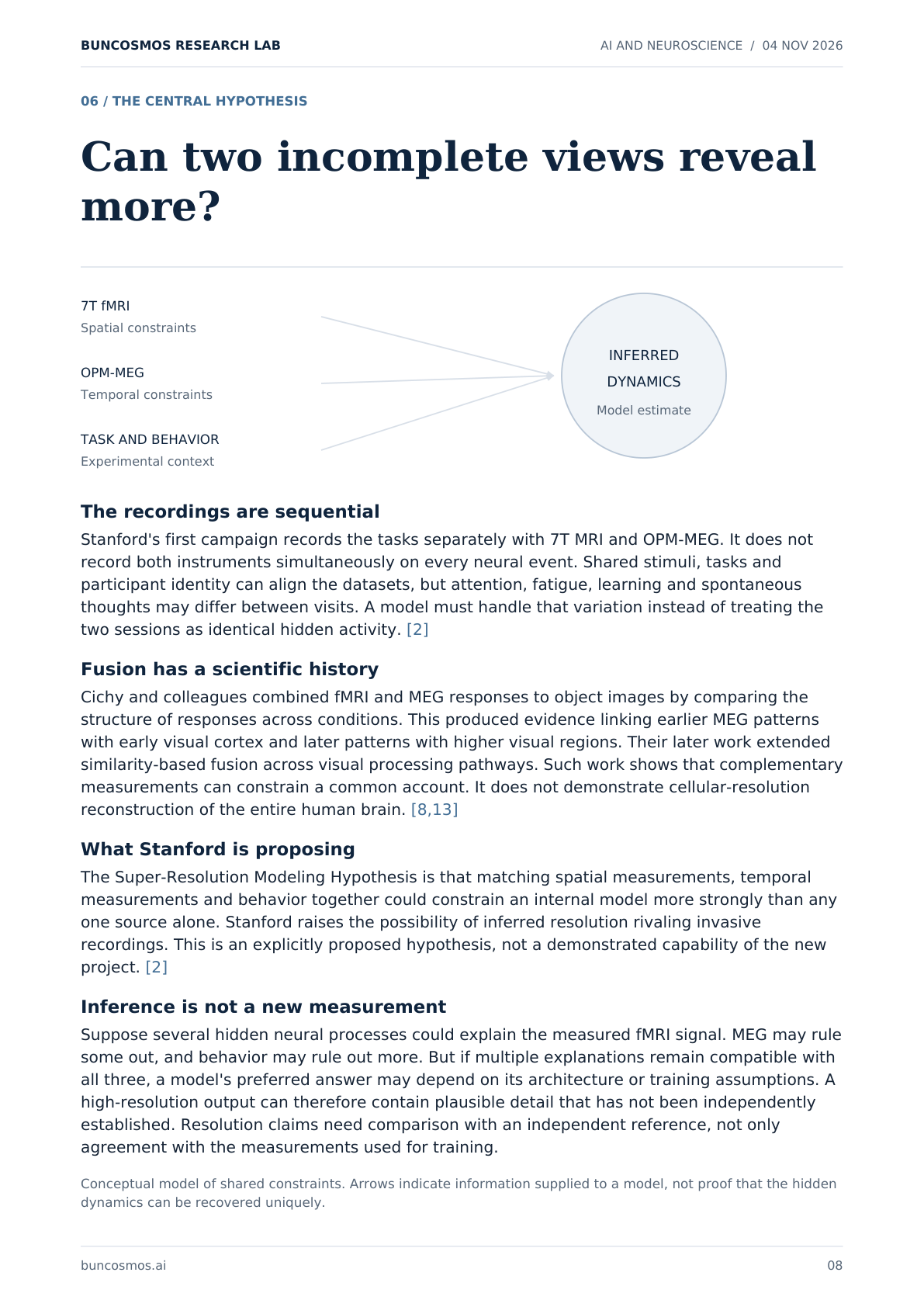
The Super-Resolution Modeling Hypothesis is that matching spatial measurements, temporal measurements and behavior together could constrain an internal model more strongly than any one source alone. Stanford raises the possibility of inferred resolution rivaling invasive recordings. This is an explicitly proposed hypothesis, not a demonstrated capability of the new project. [2]
Inference is not a new measurement
Suppose several hidden neural processes could explain the measured fMRI signal. MEG may rule some out, and behavior may rule out more. But if multiple explanations remain compatible with all three, a model's preferred answer may depend on its architecture or training assumptions. A high-resolution output can therefore contain plausible detail that has not been independently established. Resolution claims need comparison with an independent reference, not only agreement with the measurements used for training.
Conceptual model of shared constraints. Arrows indicate information supplied to a model, not proof that the hidden dynamics can be recovered uniquely.
buncosmos.ai 08
Page 9
BUNCOSMOS RESEARCH LAB AI AND NEUROSCIENCE / 04 NOV 2026
07 / FROM RECORDINGS TO MODELS
How the learning process could work
Several data streams become one learning problem
A practical pipeline would need to track the stimulus, task instructions, responses, eye movements where recorded, imaging metadata and participant identity. The learning system could then predict missing or future activity from available information. This explains how a large dataset can provide many learning examples without a human manually labeling every brain state. It is a conceptual description, not a disclosed final Stanford architecture. [3]
Self-supervision supplies targets from the data
If part of a recording is hidden, the remaining measurements can serve as input and the hidden portion as a target. If future activity is predicted, the subsequent recorded activity supplies the target. These objectives reward predictive structure, but they can also reward shortcuts. The model might use a scanner-specific pattern or recognize a repeated stimulus instead of learning a transferable cognitive relationship.
The connection to world modeling
The project website points to Counterfactual World Modeling. Bear and colleagues' 2023 preprint describes structured masking of visual data and comparisons between predictions from actual and modified inputs. Their demonstrations concern visual computations such as motion, segmentation and depth. This supplies an intellectual and technical precedent, rather than proof that the same procedure can identify causal mechanisms in a human brain. The Stanford announcement does not commit to every architectural detail of that earlier model. [2,14]
Compute is necessary but not the main evidence
Marlowe can support large training runs, with 248 H100 GPUs in the shared cluster. The reviewed materials do not establish a final parameter count, a dedicated compute allocation, training cost, required number of examples or a measured scaling law for the new brain model. More compute can optimize a chosen objective; it cannot establish that the objective measures the scientific property one wants. [9]
What scaling would need to show
A scaling study should vary useful recording hours, people, task diversity and model capacity separately where possible. It should report whether improvements continue on genuinely unseen data. More frames from the same familiar task are different evidence from transfer to a new participant or a new cognitive demand. Stanford's stated plan to determine scaling curves is therefore part of the research, not a result already in hand. [2]
buncosmos.ai 09
Page 10
BUNCOSMOS RESEARCH LAB AI AND NEUROSCIENCE / 04 NOV 2026
08 / THE EXISTING LANDSCAPE
Brain foundation models already exist
Stanford is entering an active research area. Existing models do more than classify static medical scans: some already model activity over time and predict responses to naturalistic stimuli. Its distinctive proposed contribution is the purpose-built, deeply sampled combination of modalities and cognitive tasks, rather than inventing the idea of a brain foundation model. [2,15-18]
BrainLM
Presented at ICLR 2024, BrainLM used 6,700 hours of fMRI and masked-prediction training. Its authors report brain-state forecasting, adaptation to clinical variables and transfer to external cohorts. It is a direct counterexample to the idea that all earlier foundation models merely classify anatomical images. Its particular experiments and benchmarks still do not establish a complete model of human cognition. [15]
NeuroSTORM
Cheng Wang and colleagues' peer-reviewed 2026 Nature Biomedical Engineering paper describes pretraining on 28.65 million fMRI frames from more than 50,000 people, spanning ages 5-100. It learns from four-dimensional volumes, combining three spatial dimensions with time. Its transfer tasks support general-purpose fMRI analysis; the participant count is not a measure of how deeply each individual was sampled. [16]
BrainIAC
The 2026 Nature Neuroscience paper evaluates 48,965 MRI scans across its assembled data pool. The paper specifies 32,015 scans for contrastive pretraining, so 48,965 should not be presented as the pretraining count alone. The model adapts to several clinical imaging tasks, including age estimation, tumor-related predictions and segmentation. These are valuable imaging capabilities, distinct from forecasting integrated cognitive dynamics. [17]
TRIBE v2
Meta's 2026 research describes a model combining visual, audio and language features to predict fMRI responses. Its preprint reports over 1,000 hours from 720 people and tests on new stimuli, tasks and subjects. The reviewed arXiv record is a preprint; a journal publication was not verified here. Its multimodal encoding and virtual experiments are especially relevant predecessors. Meta's release uses a noncommercial license, a reminder that available weights do not always mean unrestricted open source. [18,19]
Study sizes use different units: scans, frames, hours and people are not interchangeable. Reported benchmark improvements are not head-to-head rankings against Stanford's planned system.
buncosmos.ai 10
Page 11
BUNCOSMOS RESEARCH LAB AI AND NEUROSCIENCE / 04 NOV 2026
09 / WHAT PRIOR RESULTS SUPPORT
Useful precedents and their limits
Dense human sampling is established
The Natural Scenes Dataset collected high-resolution 7T fMRI while eight people viewed thousands of natural images across many sessions. Its publication showed that extensive individual sampling could support strong models of visual responses. That is an important precedent for spending many hours on relatively few people. Stanford's proposed multimodal campaigns expand the scope, but dense sampling itself is not new. [21]
Neural foundation models can generalize in animals
Eric Y. Wang and colleagues' 2025 Nature study recorded responses from about 135,000 neurons in 14 awake mice. A shared foundation core trained on eight mice could be adapted to others and predicted responses to stimulus types outside the natural-video training domain. This is experimentally grounded evidence that neural activity models can transfer across animals and stimuli. It concerns mouse visual cortex measured with cellular methods, not a whole human brain inferred from non-invasive recordings. [22]
Some models also address organization
A 2024 Neuron study by Margalit and colleagues, described by Stanford, used a topographic deep neural network with naturalistic input and spatial constraints. The model reproduced aspects of visual-system response organization. This is relevant because scientific usefulness can include explaining why a representation has a particular layout, rather than only minimizing prediction error. It remains a visual-system result rather than a completed general brain model. [23]
Accurate output can hide different machinery
Bertram and colleagues' 2026 preprint inspected the internal components of a neural foundation model. They reported different representational properties in its encoder, recurrent module and readout. In their analysis, high output fidelity could depend on specialized feature maps that were less biologically plausible. The paper is preliminary, but its question is directly relevant: which internal operations, if any, correspond to biological processes? [24]
What follows from these precedents
There is evidence for deep individual datasets, cross-animal transfer, multimodal prediction and some brain-like organization. None supplies a guarantee that a single integrated model will recover every cognitive mechanism. Stanford's proposal combines promising ingredients while making a larger claim that must be tested at its own scale and in its own populations.
buncosmos.ai 11
Page 12
BUNCOSMOS RESEARCH LAB AI AND NEUROSCIENCE / 04 NOV 2026
10 / EVALUATION
The tests that will decide its value
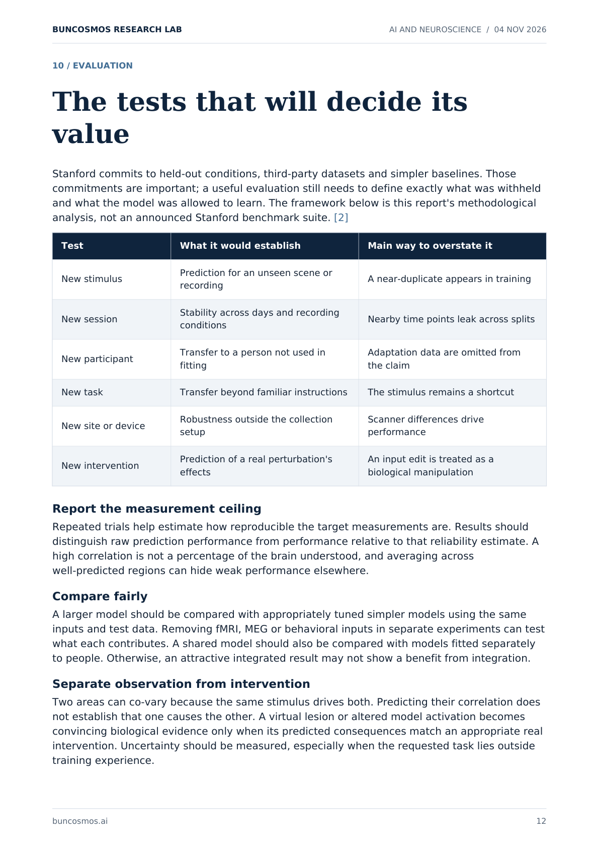
Stanford commits to held-out conditions, third-party datasets and simpler baselines. Those commitments are important; a useful evaluation still needs to define exactly what was withheld and what the model was allowed to learn. The framework below is this report's methodological analysis, not an announced Stanford benchmark suite. [2]
Test What it would establish Main way to overstate it
New stimulus Prediction for an unseen scene or recording A near-duplicate appears in training
New session Stability across days and recording conditions Nearby time points leak across splits
New participant Transfer to a person not used in fitting
Adaptation data are omitted from the claim
New task Transfer beyond familiar instructions The stimulus remains a shortcut
New site or device Robustness outside the collection setup
Scanner differences drive performance
New intervention Prediction of a real perturbation's effects
An input edit is treated as a biological manipulation
Report the measurement ceiling
Repeated trials help estimate how reproducible the target measurements are. Results should distinguish raw prediction performance from performance relative to that reliability estimate. A high correlation is not a percentage of the brain understood, and averaging across well-predicted regions can hide weak performance elsewhere.
Compare fairly
A larger model should be compared with appropriately tuned simpler models using the same inputs and test data. Removing fMRI, MEG or behavioral inputs in separate experiments can test what each contributes. A shared model should also be compared with models fitted separately to people. Otherwise, an attractive integrated result may not show a benefit from integration.
Separate observation from intervention
Two areas can co-vary because the same stimulus drives both. Predicting their correlation does not establish that one causes the other. A virtual lesion or altered model activation becomes convincing biological evidence only when its predicted consequences match an appropriate real intervention. Uncertainty should be measured, especially when the requested task lies outside training experience.
buncosmos.ai 12
Page 13
BUNCOSMOS RESEARCH LAB AI AND NEUROSCIENCE / 04 NOV 2026
11 / MEDICAL SIGNIFICANCE
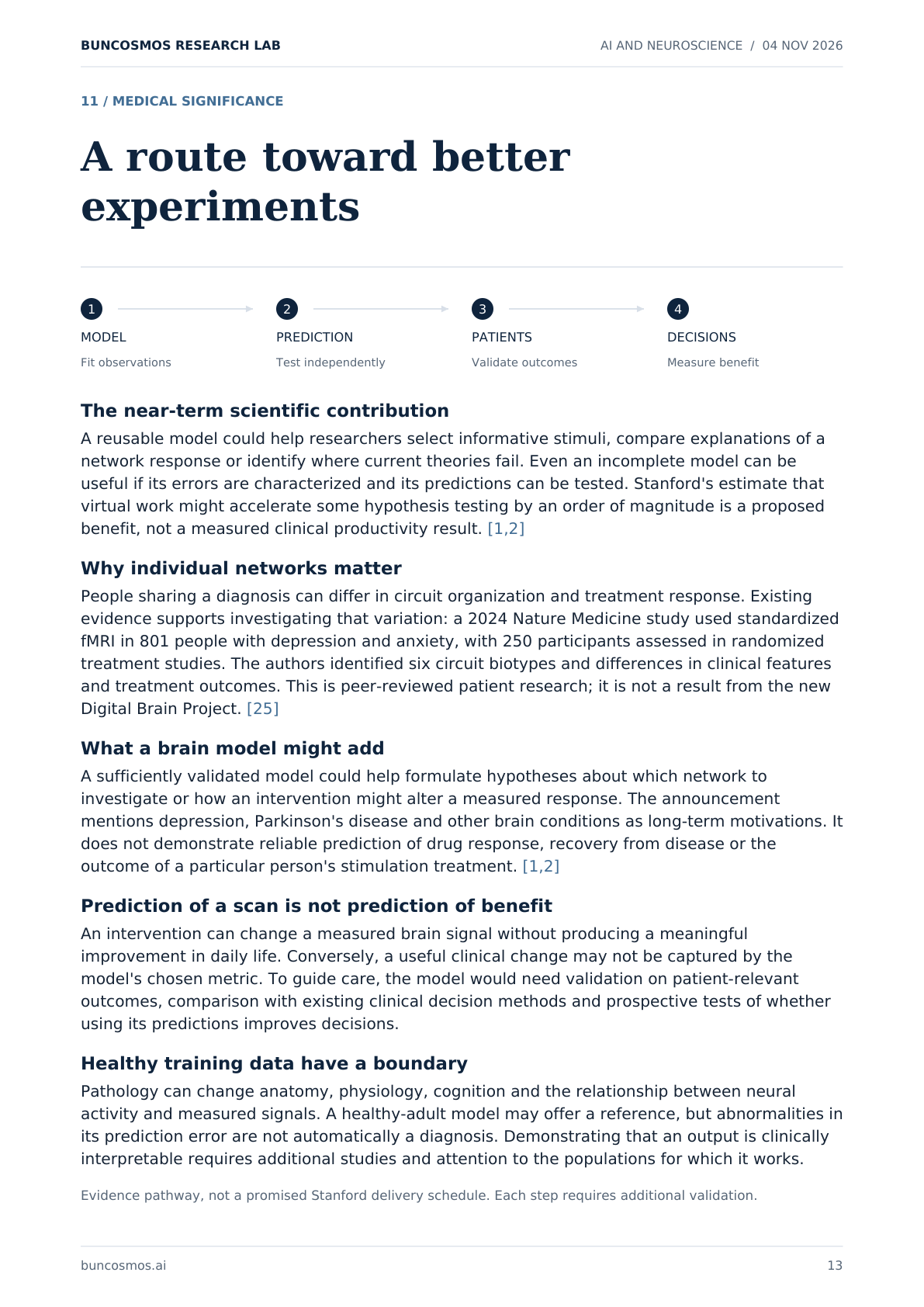
A route toward better experiments
1
2
3
4
MODEL
PREDICTION
PATIENTS
DECISIONS
Fit observations
Test independently
Validate outcomes
Measure benefit
The near-term scientific contribution
A reusable model could help researchers select informative stimuli, compare explanations of a network response or identify where current theories fail. Even an incomplete model can be useful if its errors are characterized and its predictions can be tested. Stanford's estimate that virtual work might accelerate some hypothesis testing by an order of magnitude is a proposed benefit, not a measured clinical productivity result. [1,2]
Why individual networks matter
People sharing a diagnosis can differ in circuit organization and treatment response. Existing evidence supports investigating that variation: a 2024 Nature Medicine study used standardized fMRI in 801 people with depression and anxiety, with 250 participants assessed in randomized treatment studies. The authors identified six circuit biotypes and differences in clinical features and treatment outcomes. This is peer-reviewed patient research; it is not a result from the new Digital Brain Project. [25]
What a brain model might add
A sufficiently validated model could help formulate hypotheses about which network to investigate or how an intervention might alter a measured response. The announcement mentions depression, Parkinson's disease and other brain conditions as long-term motivations. It does not demonstrate reliable prediction of drug response, recovery from disease or the outcome of a particular person's stimulation treatment. [1,2]
Prediction of a scan is not prediction of benefit
An intervention can change a measured brain signal without producing a meaningful improvement in daily life. Conversely, a useful clinical change may not be captured by the model's chosen metric. To guide care, the model would need validation on patient-relevant outcomes, comparison with existing clinical decision methods and prospective tests of whether using its predictions improves decisions.
Healthy training data have a boundary
Pathology can change anatomy, physiology, cognition and the relationship between neural activity and measured signals. A healthy-adult model may offer a reference, but abnormalities in its prediction error are not automatically a diagnosis. Demonstrating that an output is clinically interpretable requires additional studies and attention to the populations for which it works.
Evidence pathway, not a promised Stanford delivery schedule. Each step requires additional validation.
buncosmos.ai 13
Page 14
BUNCOSMOS RESEARCH LAB AI AND NEUROSCIENCE / 04 NOV 2026
12 / OPEN SCIENCE AND PRIVACY
Sharing a deeply personal dataset
Open resources are a stated commitment
Stanford describes plans for datasets, models, code, training tools, protocols and benchmarks, with public or controlled access where appropriate. The aspiration is shared scientific infrastructure. The reviewed project materials do not specify final licenses, release dates, all access criteria or the exact model artifacts that will be downloadable. Open science is a commitment to verify through releases, not evidence that everything is already available. [2]
Standards make the records usable
BIDS provides conventions for organizing imaging files and associated metadata across modalities, including fMRI and MEG. OpenNeuro provides an archive and dataset versions. These solve different problems: a standard organizes information; an archive distributes particular versions. Neither automatically guarantees that an experiment is valid or that a dataset is safe to release. [26,27]
Removing a name is not the end of privacy
Finn and colleagues showed that connectivity patterns can identify individuals within a studied group across recordings. This does not mean every public scan reveals a person's civil identity. It does show why extensive individual data deserve careful treatment. Structural images can also contain facial information; OpenNeuro's guidance requires defacing or explicit consent and ethical authorization for release without it. [27,28]
Brain decoding makes the issue concrete
Tang and colleagues' 2023 Nature Neuroscience study reconstructed aspects of continuous language from fMRI using participant-specific training. The authors found that cooperation was needed for training and application in their tests. It was not unrestricted remote mind reading. The result nevertheless demonstrates that rich neural data and learned language priors can support sensitive inferences under specific conditions. [20]
Questions a release should answer
Consent should clearly address repeated measurements, future model training and what information is shared. Governance should identify who can access potentially identifying material, how authorized use is checked and what happens when privacy risks change. Public data and controlled-access data may need different distribution paths. A model release should also explain whether it can reveal information about training participants. These are evaluation questions, not claims that Stanford has failed its safeguards.
buncosmos.ai 14
Page 15
BUNCOSMOS RESEARCH LAB AI AND NEUROSCIENCE / 04 NOV 2026
13 / PERSONAL MODELS AND CONSCIOUSNESS
A person-specific prediction is not a person
Four meanings that should remain separate
A population model captures patterns shared across participants. A person-specific model is calibrated to an individual's measured responses. A biological simulation aims to represent particular mechanisms. A reproduction of a person's mind would make a much larger claim about memories, identity and experience. Success at one level does not establish success at the next.
What personalization could mean in practice
After training a shared model, researchers could fit a smaller participant-dependent component or adjust the model with new recordings. The exact strategy remains an architectural choice, not a published Stanford implementation. The relevant tests are how much new data are needed and whether adaptation improves unseen predictions. A model that only fits a familiar person's training sessions would not demonstrate useful personalization.
Why consciousness is a separate question
Predicting a brain response and establishing subjective experience are different scientific problems. A system could predict a measurement without implementing the process responsible for that measurement in the same way. Conversely, a claim that no possible brain model could ever be conscious would require arguments beyond this project's announcement. The supported statement is that Stanford has not demonstrated a conscious digital person, and creating one is not the stated research objective. [1-3]
What consciousness research can and cannot contribute
Butlin and colleagues' peer-reviewed discussion of AI consciousness assessment emphasizes theory-informed indicators and substantial uncertainty. No single match to a brain scan is presented there as a decisive consciousness test. If future models reproduce more relevant internal mechanisms, that may motivate a separate assessment. The name digital brain, a convincing animation or a human-like conversation would not settle it. [29]
The honest interpretation of a digital twin
In this report, a digital twin means a computational predictor linked to biological measurements. It does not imply a complete replica. Every claim should specify which person, systems, tasks, signals and time scales it covers. That vocabulary allows a model to be useful without assigning it capabilities or experiences that have not been established.
buncosmos.ai 15
Page 16
BUNCOSMOS RESEARCH LAB AI AND NEUROSCIENCE / 04 NOV 2026
14 / WHAT COMES NEXT
The evidence worth watching
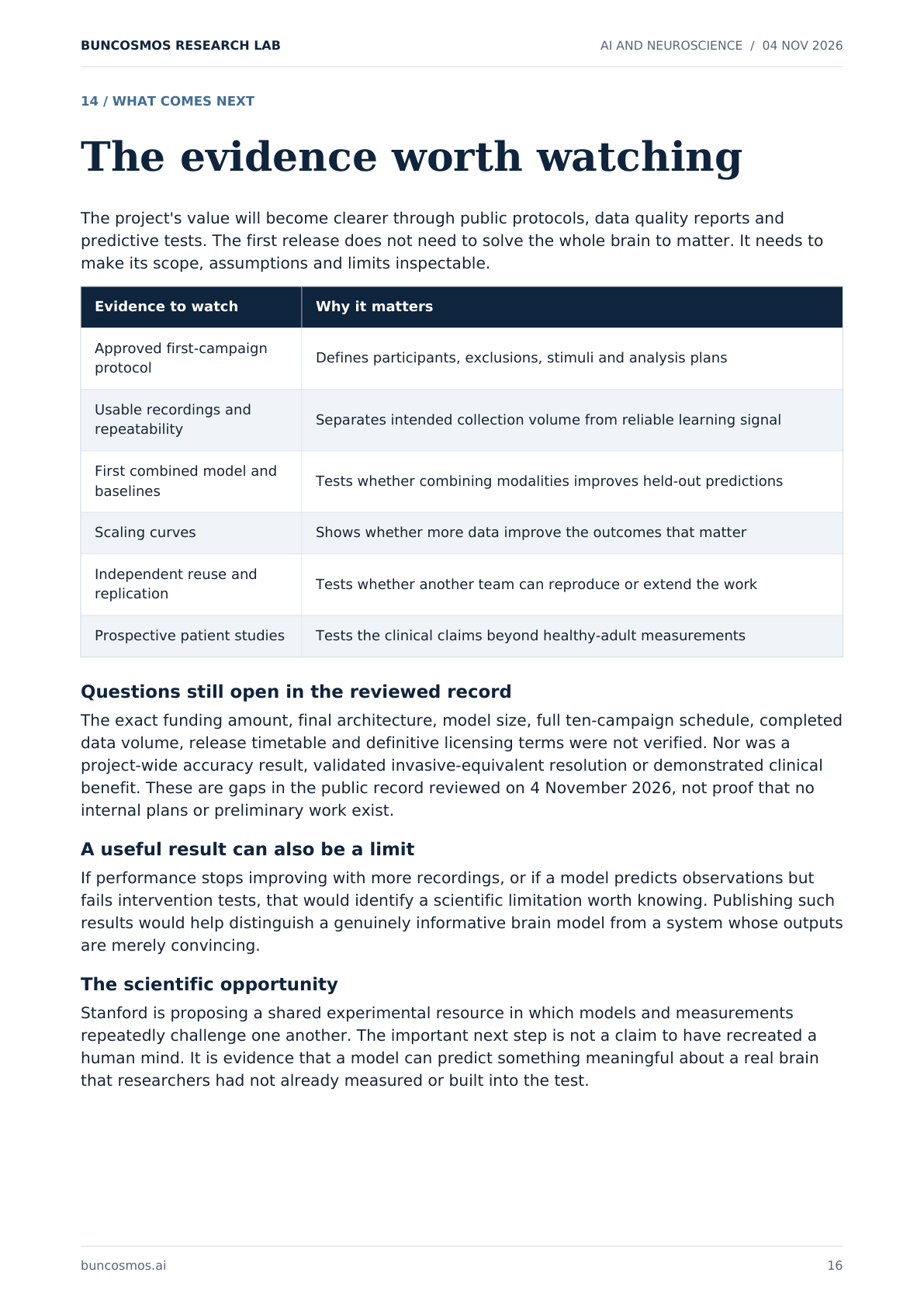
The project's value will become clearer through public protocols, data quality reports and predictive tests. The first release does not need to solve the whole brain to matter. It needs to make its scope, assumptions and limits inspectable.
Evidence to watch Why it matters
Approved first-campaign protocol Defines participants, exclusions, stimuli and analysis plans
Usable recordings and repeatability Separates intended collection volume from reliable learning signal
First combined model and baselines Tests whether combining modalities improves held-out predictions
Scaling curves Shows whether more data improve the outcomes that matter
Independent reuse and replication Tests whether another team can reproduce or extend the work
Prospective patient studies Tests the clinical claims beyond healthy-adult measurements
Questions still open in the reviewed record
The exact funding amount, final architecture, model size, full ten-campaign schedule, completed data volume, release timetable and definitive licensing terms were not verified. Nor was a project-wide accuracy result, validated invasive-equivalent resolution or demonstrated clinical benefit. These are gaps in the public record reviewed on 4 November 2026, not proof that no internal plans or preliminary work exist.
A useful result can also be a limit
If performance stops improving with more recordings, or if a model predicts observations but fails intervention tests, that would identify a scientific limitation worth knowing. Publishing such results would help distinguish a genuinely informative brain model from a system whose outputs are merely convincing.
The scientific opportunity
Stanford is proposing a shared experimental resource in which models and measurements repeatedly challenge one another. The important next step is not a claim to have recreated a human mind. It is evidence that a model can predict something meaningful about a real brain that researchers had not already measured or built into the test.
buncosmos.ai 16
Page 17
BUNCOSMOS RESEARCH LAB AI AND NEUROSCIENCE / 04 NOV 2026
READER REFERENCE
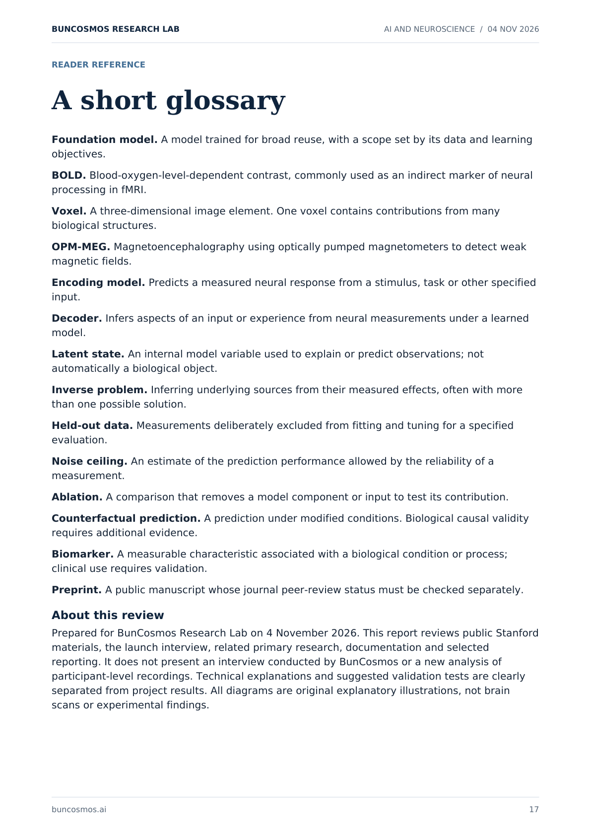
A short glossary
Foundation model. A model trained for broad reuse, with a scope set by its data and learning objectives.
BOLD. Blood-oxygen-level-dependent contrast, commonly used as an indirect marker of neural processing in fMRI.
Voxel. A three-dimensional image element. One voxel contains contributions from many biological structures.
OPM-MEG. Magnetoencephalography using optically pumped magnetometers to detect weak magnetic fields.
Encoding model. Predicts a measured neural response from a stimulus, task or other specified input.
Decoder. Infers aspects of an input or experience from neural measurements under a learned model.
Latent state. An internal model variable used to explain or predict observations; not automatically a biological object.
Inverse problem. Inferring underlying sources from their measured effects, often with more than one possible solution.
Held-out data. Measurements deliberately excluded from fitting and tuning for a specified evaluation.
Noise ceiling. An estimate of the prediction performance allowed by the reliability of a measurement.
Ablation. A comparison that removes a model component or input to test its contribution.
Counterfactual prediction. A prediction under modified conditions. Biological causal validity requires additional evidence.
Biomarker. A measurable characteristic associated with a biological condition or process; clinical use requires validation.
Preprint. A public manuscript whose journal peer-review status must be checked separately.
About this review
Prepared for BunCosmos Research Lab on 4 November 2026. This report reviews public Stanford materials, the launch interview, related primary research, documentation and selected reporting. It does not present an interview conducted by BunCosmos or a new analysis of participant-level recordings. Technical explanations and suggested validation tests are clearly separated from project results. All diagrams are original explanatory illustrations, not brain scans or experimental findings.
buncosmos.ai 17
Page 18
BUNCOSMOS RESEARCH LAB AI AND NEUROSCIENCE / 04 NOV 2026
SOURCES / PRIMARY RESEARCH AND PROJECT RECORDS
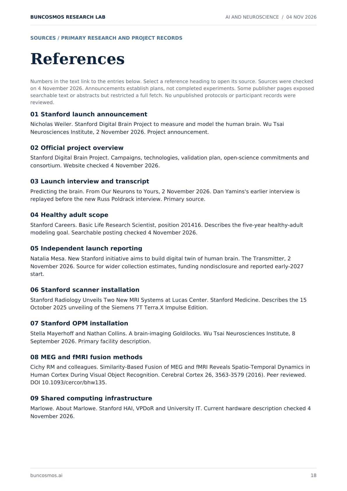
References
Numbers in the text link to the entries below. Select a reference heading to open its source. Sources were checked on 4 November 2026. Announcements establish plans, not completed experiments. Some publisher pages exposed searchable text or abstracts but restricted a full fetch. No unpublished protocols or participant records were reviewed.
01 Stanford launch announcement
Nicholas Weiler. Stanford Digital Brain Project to measure and model the human brain. Wu Tsai Neurosciences Institute, 2 November 2026. Project announcement.
02 Official project overview
Stanford Digital Brain Project. Campaigns, technologies, validation plan, open-science commitments and consortium. Website checked 4 November 2026.
03 Launch interview and transcript
Predicting the brain. From Our Neurons to Yours, 2 November 2026. Dan Yamins's earlier interview is replayed before the new Russ Poldrack interview. Primary source.
04 Healthy adult scope
Stanford Careers. Basic Life Research Scientist, position 201416. Describes the five-year healthy-adult modeling goal. Searchable posting checked 4 November 2026.
05 Independent launch reporting
Natalia Mesa. New Stanford initiative aims to build digital twin of human brain. The Transmitter, 2 November 2026. Source for wider collection estimates, funding nondisclosure and reported early-2027 start.
06 Stanford scanner installation
Stanford Radiology Unveils Two New MRI Systems at Lucas Center. Stanford Medicine. Describes the 15 October 2025 unveiling of the Siemens 7T Terra.X Impulse Edition.
07 Stanford OPM installation
Stella Mayerhoff and Nathan Collins. A brain-imaging Goldilocks. Wu Tsai Neurosciences Institute, 8 September 2026. Primary facility description.
08 MEG and fMRI fusion methods
Cichy RM and colleagues. Similarity-Based Fusion of MEG and fMRI Reveals Spatio-Temporal Dynamics in Human Cortex During Visual Object Recognition. Cerebral Cortex 26, 3563-3579 (2016). Peer reviewed. DOI 10.1093/cercor/bhw135.
09 Shared computing infrastructure
Marlowe. About Marlowe. Stanford HAI, VPDoR and University IT. Current hardware description checked 4 November 2026.
buncosmos.ai 18
Page 19
BUNCOSMOS RESEARCH LAB AI AND NEUROSCIENCE / 04 NOV 2026
SOURCES / CONTINUED
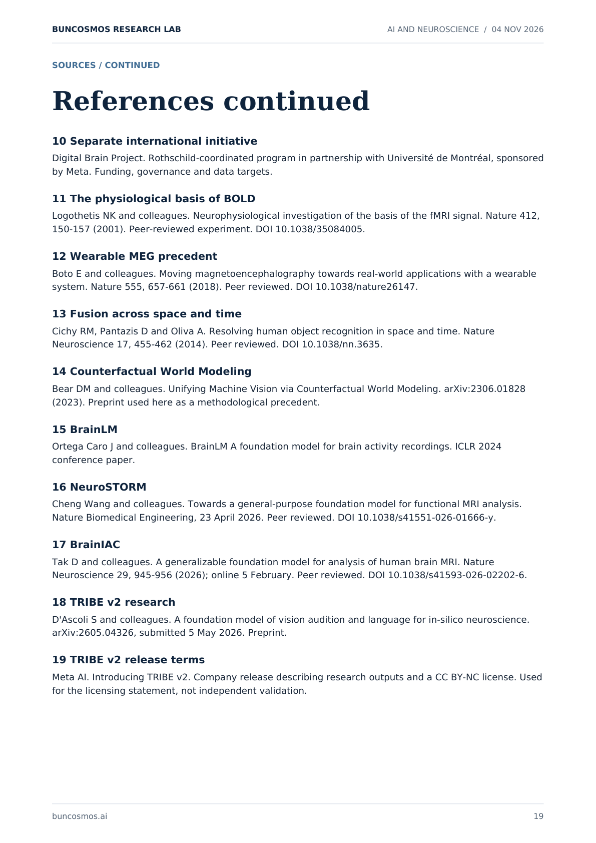
References continued
10 Separate international initiative
Digital Brain Project. Rothschild-coordinated program in partnership with Université de Montréal, sponsored by Meta. Funding, governance and data targets.
11 The physiological basis of BOLD
Logothetis NK and colleagues. Neurophysiological investigation of the basis of the fMRI signal. Nature 412, 150-157 (2001). Peer-reviewed experiment. DOI 10.1038/35084005.
12 Wearable MEG precedent
Boto E and colleagues. Moving magnetoencephalography towards real-world applications with a wearable system. Nature 555, 657-661 (2018). Peer reviewed. DOI 10.1038/nature26147.
13 Fusion across space and time
Cichy RM, Pantazis D and Oliva A. Resolving human object recognition in space and time. Nature Neuroscience 17, 455-462 (2014). Peer reviewed. DOI 10.1038/nn.3635.
14 Counterfactual World Modeling
Bear DM and colleagues. Unifying Machine Vision via Counterfactual World Modeling. arXiv:2306.01828 (2023). Preprint used here as a methodological precedent.
15 BrainLM
Ortega Caro J and colleagues. BrainLM A foundation model for brain activity recordings. ICLR 2024 conference paper.
16 NeuroSTORM
Cheng Wang and colleagues. Towards a general-purpose foundation model for functional MRI analysis. Nature Biomedical Engineering, 23 April 2026. Peer reviewed. DOI 10.1038/s41551-026-01666-y.
17 BrainIAC
Tak D and colleagues. A generalizable foundation model for analysis of human brain MRI. Nature Neuroscience 29, 945-956 (2026); online 5 February. Peer reviewed. DOI 10.1038/s41593-026-02202-6.
18 TRIBE v2 research
D'Ascoli S and colleagues. A foundation model of vision audition and language for in-silico neuroscience. arXiv:2605.04326, submitted 5 May 2026. Preprint.
19 TRIBE v2 release terms
Meta AI. Introducing TRIBE v2. Company release describing research outputs and a CC BY-NC license. Used for the licensing statement, not independent validation.
buncosmos.ai 19
Page 20
BUNCOSMOS RESEARCH LAB AI AND NEUROSCIENCE / 04 NOV 2026
SOURCES / CONTINUED
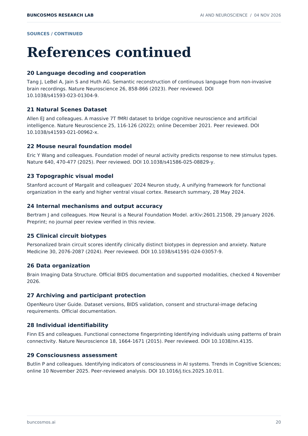
References continued
20 Language decoding and cooperation
Tang J, LeBel A, Jain S and Huth AG. Semantic reconstruction of continuous language from non-invasive brain recordings. Nature Neuroscience 26, 858-866 (2023). Peer reviewed. DOI 10.1038/s41593-023-01304-9.
21 Natural Scenes Dataset
Allen EJ and colleagues. A massive 7T fMRI dataset to bridge cognitive neuroscience and artificial intelligence. Nature Neuroscience 25, 116-126 (2022); online December 2021. Peer reviewed. DOI 10.1038/s41593-021-00962-x.
22 Mouse neural foundation model
Eric Y Wang and colleagues. Foundation model of neural activity predicts response to new stimulus types. Nature 640, 470-477 (2025). Peer reviewed. DOI 10.1038/s41586-025-08829-y.
23 Topographic visual model
Stanford account of Margalit and colleagues' 2024 Neuron study, A unifying framework for functional organization in the early and higher ventral visual cortex. Research summary, 28 May 2024.
24 Internal mechanisms and output accuracy
Bertram J and colleagues. How Neural is a Neural Foundation Model. arXiv:2601.21508, 29 January 2026. Preprint; no journal peer review verified in this review.
25 Clinical circuit biotypes
Personalized brain circuit scores identify clinically distinct biotypes in depression and anxiety. Nature Medicine 30, 2076-2087 (2024). Peer reviewed. DOI 10.1038/s41591-024-03057-9.
26 Data organization
Brain Imaging Data Structure. Official BIDS documentation and supported modalities, checked 4 November 2026.
27 Archiving and participant protection
OpenNeuro User Guide. Dataset versions, BIDS validation, consent and structural-image defacing requirements. Official documentation.
28 Individual identifiability
Finn ES and colleagues. Functional connectome fingerprinting Identifying individuals using patterns of brain connectivity. Nature Neuroscience 18, 1664-1671 (2015). Peer reviewed. DOI 10.1038/nn.4135.
29 Consciousness assessment
Butlin P and colleagues. Identifying indicators of consciousness in AI systems. Trends in Cognitive Sciences; online 10 November 2025. Peer-reviewed analysis. DOI 10.1016/j.tics.2025.10.011.
buncosmos.ai 20